It is Suitable For
· Those in Tokyo looking for a detailed liver scan using advanced technology.
· Those with liver problems such as hepatitis.
· Those with suspected liver cancer.
· Those who want a clear diagnosis of liver treatment.
· Those who wish to monitor how well their liver treatment is working.
What Is Liver Contrast MRI And Tumour Marker?
Liver Contrast MRI is a type of scan that helps the doctor see the liver more clearly by using strong magnets, radio waves, and a specific dye called a contrast agent, usually gadolinium. This dye will be injected to the vein to help highlight blood vessels, bile ducts, and liver tissues in the images. The scan shows detailed pictures that help to detect liver tumours, check for liver disease, analyze blood flow, and see how well liver treatments are working. It is especially helpful in spotting liver cancer, such as hepatocellular carcinoma (HCC), and in telling the difference between cancerous and non-cancerous growths.
Liver Tumour Marker is studied by substance, often proteins, produced either by the liver or by the tumours within the liver. One important marker is alpha-fetoprotein (AFP), which can be found in significantly high amounts within the blood when someone has liver cancer. The doctor uses the markers to help detect liver cancer early, especially in people with liver problems like hepatitis. While tumour markers alone cannot confirm cancer, they are useful when combined with scans like MRI to track diseases and see how well treatments are working.
How It Works
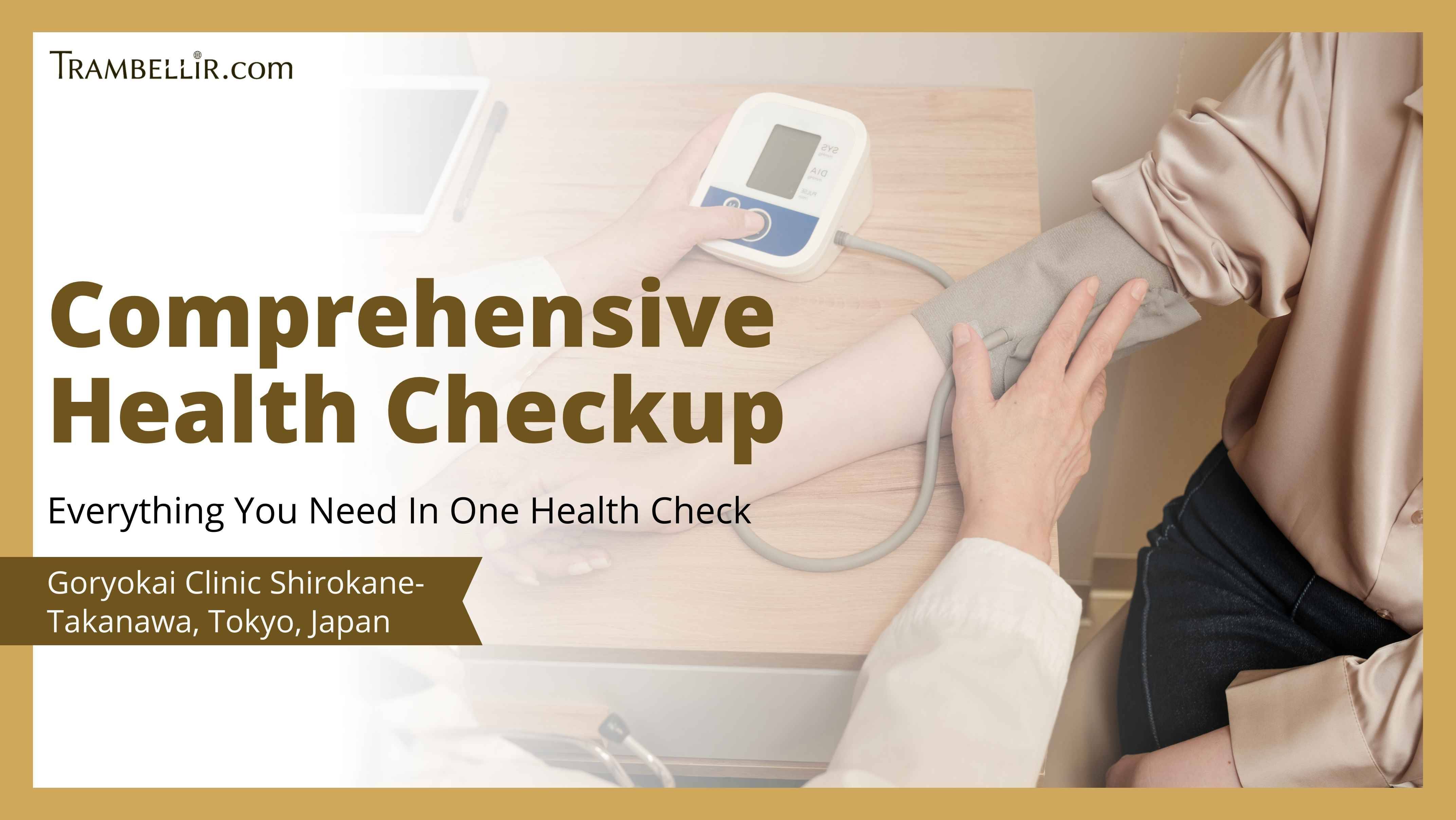
A Liver Contrast MRI is a non-invasive imaging technique that uses strong magnetic fields and radiofrequency waves to produce high-resolution, cross-sectional images of the liver. Prior to the scan, a contrast agent, typically gadolinium based, is administered intravenously. This contrast enhances the visibility of liver tissues, blood vessels, and bile ducts, helping to differentiate between normal and abnormal structures. During the procedure, patients will lie on a motorized table that slides into a cylindrical MRI scanner. Patients are asked to remain still and may occasionally be instructed to hold their breath for a few seconds to minimize motion and improve image clarity. The resulting images will be interpreted by the doctor, who assesses for liver tumours, vascular abnormalities, biliary disease, and other hepatic pathology.
Meanwhile, Tumour Markers measure substances such as proteins that are most commonly found in the blood which may help to indicate the presence of liver cancer. The most widely used marker is alpha-fetoprotein (AFP); elevated AFP levels are often associated with hepatocellular carcinoma (HCC), though the increase in number may also occur in chronic liver conditions such as hepatitis or cirrhosis. The test involves collecting a blood sample, which will be analyzed in a laboratory to determine the concentration of specific markers. While tumour markers alone are not diagnostic, they serve as valuable tools when used alongside imaging studies such as MRI. AFP and other markers are used in screening high-risk patients, assisting in diagnosis and tracking disease progression or treatment response.
Liver Contrast MRI And Tumour Marker Procedure
Liver Contrast MRI
1. Consultation will be conducted by the doctor.
2. All metal objects like jewelry, hairpins, or watches will be removed.
3. The doctor will administer a gadolinium-based contrast agent to the patient’s arm.
4. Patients will lie flat on a table that moves into the MRI machine.
5. Earplugs will be provided as the machine will make loud noise.
6. Results will be reviewed by the doctor to determine if further testing is required.
Tumour Marker
1. Consultation will be conducted by the doctor.
2. A blood sample will be drawn.
3. The blood sample will be sent to the lab to test for tumour markers.
4. Results will be reviewed by the doctor to determine if further testing is required.
Advantages and Limitations of Liver Contrast MRI And Tumour Marker
<Advantages>
· Shows very detailed images of the liver and blood vessels
· Helps detect tumours, blockage, or the liver problems early
· Useful for staging cancer and tracking treatment progress
<Limitations>
· Not suitable for those with certain metal implants
· May required follow up test to confirm findings
Liver Contrast MRI And Tumour Marker Preparation
· Inform the doctor for any ongoing medications or allergies
· Avoid eating and drinking a few hours before taking the test
· Remove all metal items from body before the taking the test
Liver Contrast MRI And Tumour Marker Post Care
· Ensure to drink enough water to stay hydrated after the test
· Patients can return to work or daily activities after the test
FAQ
Q1: Is Liver Contrast MRI And Tumour Marker painful?
A1: Liver Contrast MRI And Tumour Marker is non-invasive and painless.
Q2: How long does Liver Contrast MRI And Tumour Marker procedure take?
A2: Liver Contrast MRI And Tumour Marker procedure takes around 60 to 90 minutes.
Q3: Is the contrast dye used during Liver Contrast MRI And Tumour Marker safe?
A3: Yes, the contrast dye used during Liver Contrast MRI And Tumour Marker are generally safe to be used on everyone, however in rare cases, allergies may occur and shall be addressed immediately.
Q4: How long do I have to wait to get Liver Contrast MRI And Tumour Marker results?
A4: The results for Liver Contrast MRI And Tumour Marker may take 7 to 14 days.
Q5: Is there any downtime after Liver Contrast MRI And Tumour Marker?
A5: There is no downtime after Liver Contrast MRI And Tumour Marker, patients may resume to their normal activities after the test.
Clinic Information
[Clinic Name] Forest of Regeneration Clinic Roppongi (Saisei No Mori Clinic)
[Location] Tokyo, Japan
[Nearest Station/ Nearest Tourist Destination] Clinic is located about 5 mins away from The National Art Center and 9 mins Roppngi Tunnel Wallart
[Clinic Opening Time] Monday - Friday (10:00AM - 3:00PM) ; Saturday (10:00AM - 2:00PM) ; Clinic Closed on Sunday, Public Holidays and every 1st, 3rd and 5th Fridays
| Operation Time |
60 - 90 mins
|
| Recovery Time |
- |
| Number of Postoperative Visit |
None |
| Hospitalization |
Not required |
| Limitations |
None |
| Cancellation Policy |
No charge for cancellations made at least 48 hrs prior appointment date* |
| Include In This Price |
Consultation Fee, Interpreter Fee, Tax |
| Location |
M's BILD. 4F-A, 7-10-1 Roppongi, Minato-ku, Tokyo 106-0032 |
Terms of Service
Trambellir is a health tourism platform and does not in any way provide, promote, or sell any
medication or medical treatment to the Customers.
Customers are advised to contact the healthcare providers directly for any enquiries related to
medical treatments.